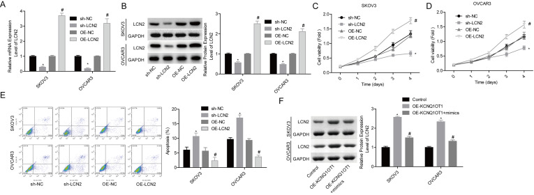
Figure 6.
Upregulation of LCN2 increased cell proliferation and inhibited cell apoptosis in ovarian cancer. (A, B) qPCR and Western blot assays were performed to examine LCN2 expression levels after 48 h of cell transfection with OE-LCN2, OE-NC, sh-LCN2 or sh-NC in SKOV3 and OVCAR3 cells (*p < 0.05, sh-LCN2 group vs. sh-NC group; #p < 0.05, OE-LCN2 group vs. OE-NC group). (C, D) MTT assay was carried out to detect cell proliferation (*p < 0.05, sh-LCN2 group vs. sh-NC group; #p < 0.05, OE-LCN2 group vs. OE-NC group). (E) Flow cytometry assay was used to detect cell apoptosis (*p < 0.05, sh-LCN2 group vs. sh-NC group; #p < 0.05, OE-LCN2 group vs. OE-NC group). (F) Western blot assay was used to detect the protein expression level of LCN2 after SKOV3 and OVCAR3 cells were transfected with OE-KCNQ1OT1, OE- KCNQ1OT1 + mimics, or the control vectors (*p < 0.05, OE-KCNQ1OT1 group vs. control group; #p < 0.05, OE-KCNQ1OT1 + mimic group vs. OE-KCNQ1OT1 group).

