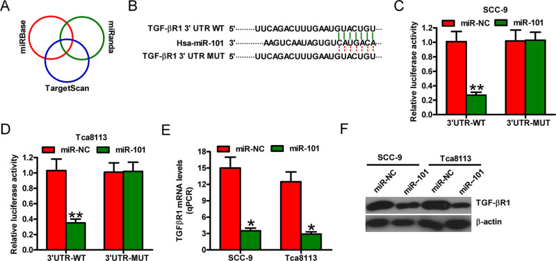
Figure 2.
TGF-βR1 is a direct target of miR-101. (A) Prediction of the potential targets of miR-101 by integrating the results of three algorithms (TargetScan, miRBase, and miRanda). (B) The 3′-untranslated region (3′-UTR) of TGF-βR1 was predicted to contain a complementary region of miR-101 seed sequences. (C, D) Luciferase reporter plasmids harboring the WT or MUT 3′-UTR of TGF-βR1 were cotransfected with miR-NC or miR-101 mimics into the SCC-9 or Tca8113 cells. Luciferase reporter assays were performed at 24 h after cotransfection. The normalized luciferase activity in the group (miR-NC + the empty plasmid) was set to 1. The (E) mRNA and (F) protein levels of TGF-βR1 were determined by qPCR and Western blot assays in SCC-9 and Tca8113 cells transfected with miR-NC or miR-101. Data are presented as the mean ± SD of three replicates. *p < 0.05 versus miR-NC group; **p < 0.01 versus miR-NC group. MUT, mutate; NC, negative control; WT, wild type.

