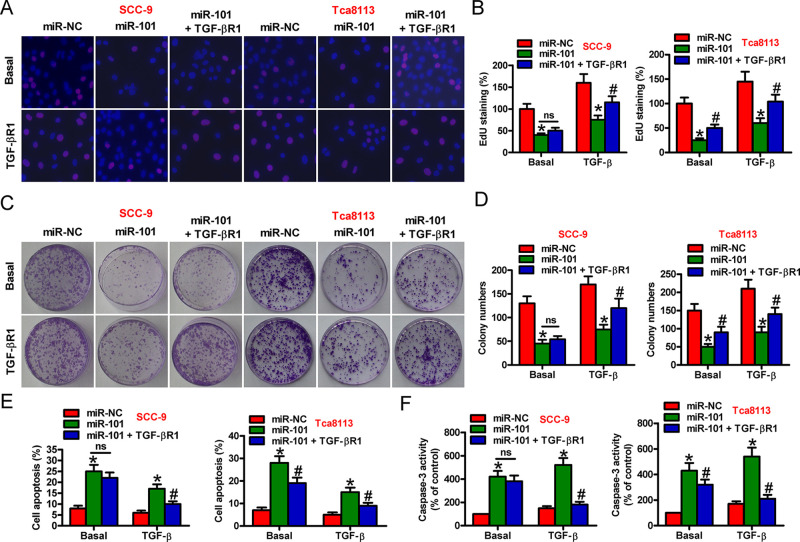
Figure 3.
Suppression of TGF-β1-induced proliferation and apoptosis resistance by miR-101 was mediated by TGF-βR1 in OSCC cells. SCC-9 and Tca8113 cells were transfected with miR-NC, miR-101, or miR-101 + TGF-βR1-expressing plasmid in the absence or presence of TGF-β1. (A) EdU assay was performed to evaluate cell proliferation. (B) Percentage of EdU-positive cells in (A). (C) Colony formation assay was conducted to assess cell proliferation. (D) The number of colonies in (C) was counted. (E) Flow cytometry assay was performed to detect the apoptotic cells. Apoptotic rate was calculated. (F) Caspase 3 activity was measured to reflect cell apoptosis. Data are expressed as the mean ± SD of three separate experiments. *p < 0.05 versus miR-NC group; #p < 0.05 versus miR-101 group. ns, not significant.

