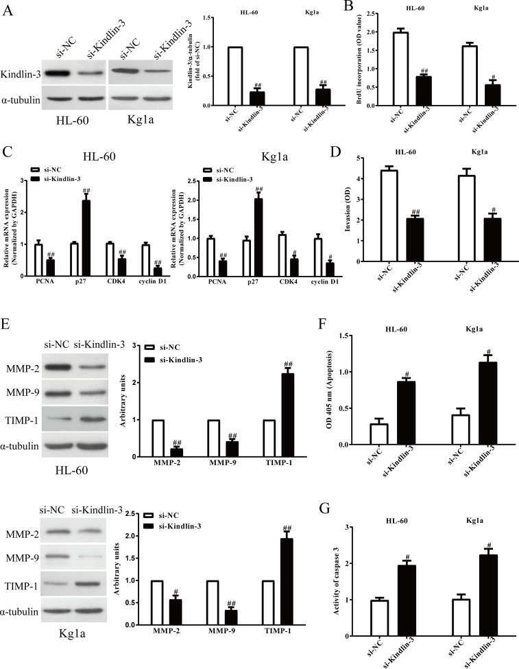
Figure 5.
The effects of kindlin-3 silencing on cell proliferation, invasion, and epithelial–mesenchymal transition (EMT) in AML cells. HL-60 and Kg1a cells were transfected with si-kindlin-3 or si-NC for 48 h. (A) The protein expressions of kindlin-3 were determined by Western blot assay. (B) Cell proliferation was assessed by BrdU-ELISA assay. (C) The mRNA expressions of PCNA, CDK4, cyclin D1, and p27 were determined by qRT-PCR. (D) The invasion was assessed by Transwell assay. (E) The protein expressions of MMP-2, MMP-9, and TIMP-1 were detected by Western blot assay. (F) Cell apoptosis was measured by nucleosomal degradation by using Roche’s cell death ELISA detection kit. (G) The activities of caspase 3 were determined by caspase 3 activity detection assay. All data are presented as mean ± SEM, n = 6. #p < 0.05, ##p < 0.01 versus si-NC.

