Figure 5.
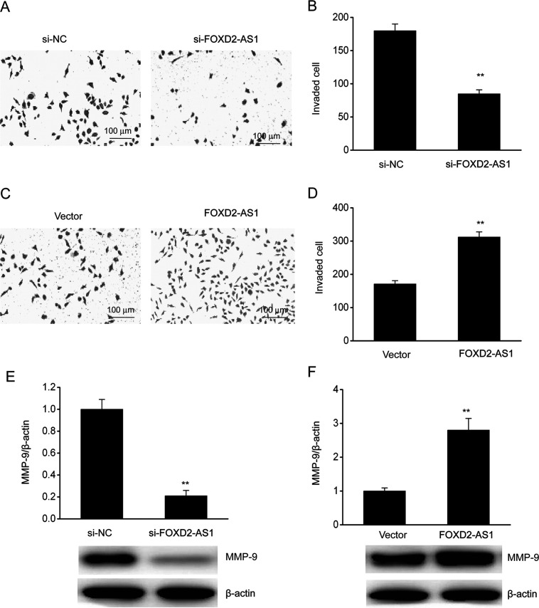
FOXD2-AS1 promoted the invasion of ESCC cells. (A) A Transwell assay was performed to evaluate invasion in TE-1/DDP cells. (B) Quantitative assessment of invading TE-1/DDP cells transfected with FOXD2-AS1 siRNA (si-FOXD2-AS1) or negative control siRNA (si-NC). (C) A Transwell assay was performed to evaluate invasion in TE-1/DDP cells. (D) Quantitative assessment of invading TE-1/DDP cells transfected with the FOXD2-AS1 expression vector (FOXD2-AS1) or a control vector (vector). (E) Representative immunoblot and quantitative evaluation of MMP-9 in TE-1/DDP cells transfected with si-FOXD2-AS1 or si-NC. (F) Representative immunoblot and quantitative evaluation of MMP-9 in TE-1/DDP cells transfected with FOXD2-AS1 or the control vector. The results are shown as the means ± SEM. **p < 0.01 versus the NC vector group.

