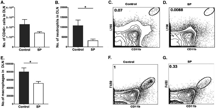
FIG 4.
Mice fed SP presented diminished innate inflammatory responses in ocular DLN tissue samples. Orally SP-fed and control C57BL/6 mice were infected with HSV-1, and FACS analysis was performed after 15 days p.i. (A) Total numbers of leukocytes in the DLN. (B) Numbers of neutrophils accumulating in the DLN. (C, D) FACS plots showing the frequencies of neutrophils (CD45+ CD11b+ Ly6G+) in the DLN. (E) Numbers of macrophages in the DLN. (F, G) FACS plots revealing frequencies of macrophages (CD45+ CD11b+ F4/80+) in the DLN. Gating for neutrophils and macrophages in the DLN was carried out as described elsewhere (59, 60). Data represent mean results ± SEM. All data were analyzed by an unpaired Student t test, and significance levels were determined (*, P ≤ 0.05; **, P ≤ 0.01; ***, P ≤ 0.001; ****, P ≤ 0.0001).

