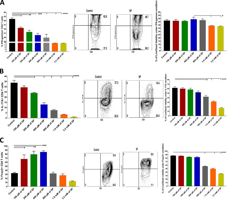
FIG 9.
Effects of addition of SP to induction cultures for Th1 cells, Th17 cells, and Treg. Splenocytes from RAG2–/– mice were cultured ex vivo in the presence of 1 μg/ml of anti-CD3/CD28 Abs as well as rIL-2 (100 U/ml) and TGF-β (0.5 ng/ml) for Treg induction, IL-12 (5 to 10 ng/ml) and anti-IL-4 (10 μg/ml) for Th1 induction, and IL-6 (25 ng/ml) and TGF-β (1 ng/ml) for Th17 induction, along with various concentrations of SP (100 μM to 3.2 mM). After 5 days, cells were harvested and analyzed for the expression of the respective immune phenotype. (A to C) Histogram analysis to measure the effects of SP on the differentiation of Th1 cells (CD4+ IFN-γ+) (A), Th17 cells (CD4+ IL-17A+) (B), and Treg (CD4+ Foxp3+) (C) and on their viability, presented as bar graphs (left) and FACS plots (right). For Th1 and Th17 cell enumeration, stimulation with PMA-ionomycin was performed for 4 h. Data represent mean results ± SEM. All data were analyzed by ANOVA, and significance levels were determined (*, P ≤ 0.05; **, P ≤ 0.01; ***, P ≤ 0.001; ****, P ≤ 0.0001).

