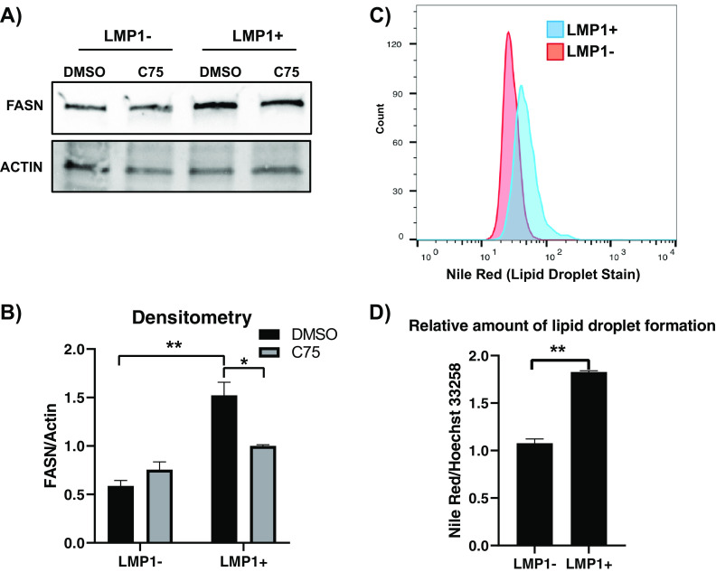
FIG 2.
LMP1 leads to increased FASN and lipid droplet formation. (A) Western blot of the EBV-negative B-cell line DG75 transduced with retroviral particles containing either pBABE (empty vector) or pBABE-HA-LMP1 vectors and treated with 10 µg/ml the FASN inhibitor C75 for 24 h. Cell lines were probed for FASN. Actin served as a loading control. (B) Densitometry of FASN/actin normalized to untreated empty vector (pBABE). (C) FACS analysis of Nile red fluorescence staining (excitation, 385 nm; emission, 535 nm) for lipid droplets in the DG75 cell line transfected with an empty plasmid vector or LMP1 expression construct. (D) The relative amount of lipid droplet formation was calculated by plate reader by normalizing the Hoechst 33342 fluorescence (excitation, 355 nm; emission, 460 nm) to the Nile red signal in each well. Error bars represent standard deviations from two independent experiments. P values for significant differences (Student's t test) are summarized by two asterisks (P < 0.01) or one asterisk (P < 0.05).

