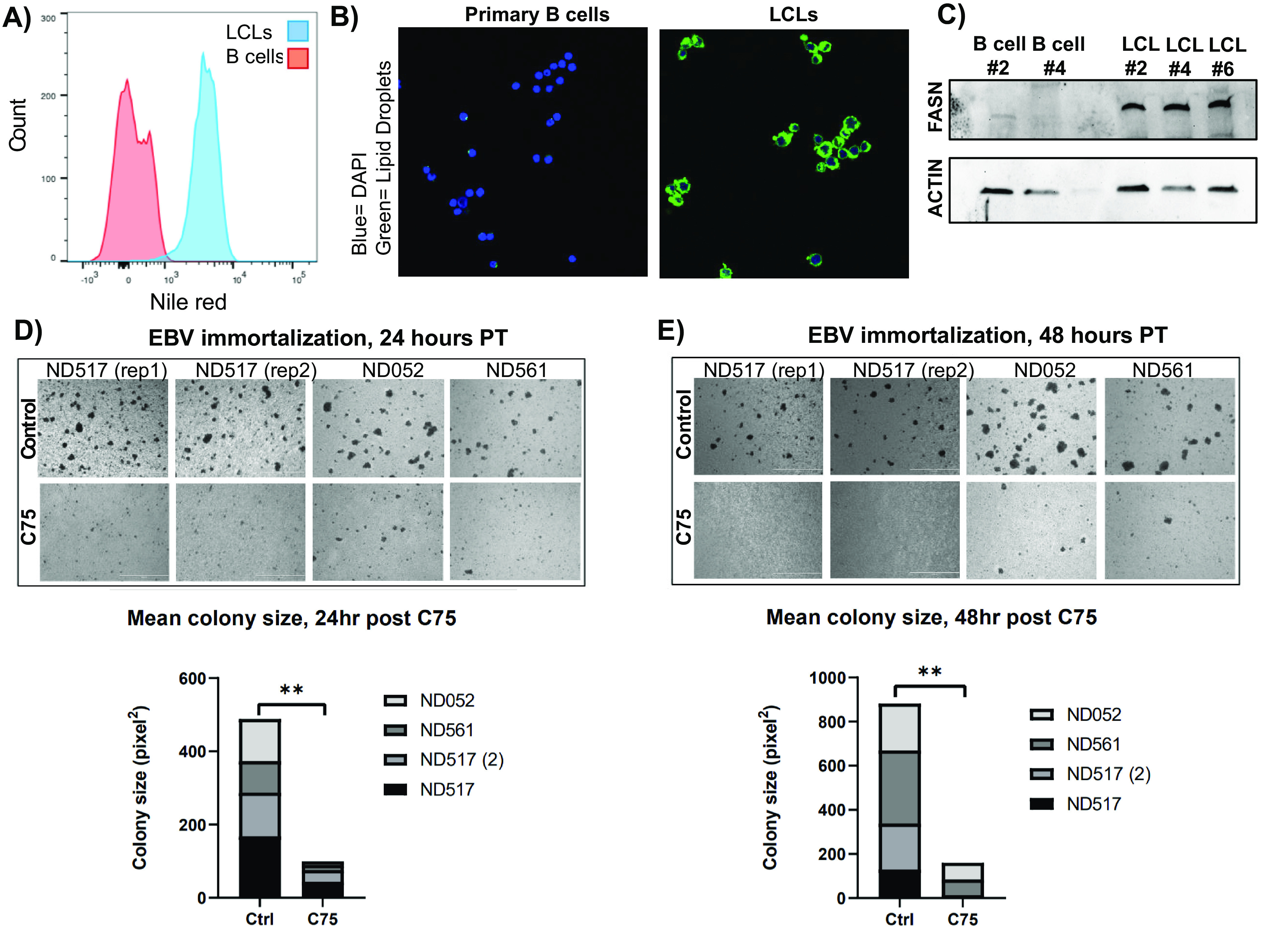
FIG 4.

EBV-induced immortalization of B cells upregulates FASN and lipogenesis. (A) FACS analysis of Nile red fluorescence staining (excitation, 385 nm; emission, 535 nm) for lipid droplets overlaying primary B cells with LCLs. (B) Confocal microscopy of Nile red fluorescence staining (excitation, 385 nm; emission, 535 nm) for lipid droplets in primary B cells and LCLs. Cells were counterstained with 4′,6-diamidino-2-phenylindole (DAPI) to stain cell nuclei. (C) Western blot for FASN in primary B cells and their matched LCLs. Actin served as a loading control. (D and E) Imaging of primary B-cell EBV immortalization. Ten million cells per group were collected from three donors (one donor was assayed at two independent times) and infected with B95.8 strain EBV 24 h prior to treatment. Cells were imaged on a Nikon TE2000 inverted microscope at ×4 magnification 24 (D) and 48 (E) h after C75 treatment. Statistics for average colony size were collected using the “analyze particle” feature of ImageJ for 30 randomized, nonoverlapping images taken of each group. The 30 mean colony size values were then averaged. P values for significant differences (Student's t test) are summarized by three asterisks (P < 0.001), two asterisks (P < 0.01), or one asterisk (P < 0.05).
