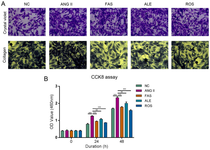
Figure 2.
ALE, FAS and ROS therapy alleviate the process of Ang II-promoted cell fibrosis and viability of cardiac fibroblasts. (A) Crystal violet staining (magnification, x200) shows cells were proliferative and growth was dense in the Ang II group. There was no difference in cell morphology between the ALE and Ang II groups. The FAS and ROS groups had fewer cells, more cytoplasm, and some cells were round compared with those in Ang II group. Collagen staining (magnification, x200) in the Ang II group was more intense compared with that in the NC group. ALE, FAS, and ROS groups was weak compared with the Ang II group. (B) Cell proliferation assay shows Ang II significantly promote proliferation compared with NC group, whilst the rates of proliferation in the ROS, ALE, and FAS treatment groups were lower compared Ang II group at both 24 and 48 h. **P<0.01 and ***P<0.001. NC, negative control; Ang II, angiotensin II; ROS, Ang II + rosuvastatin; ALE, Ang II + alendronate; FAS, Ang II + fasudil.

