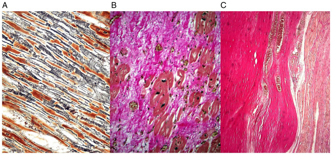
Figure 1.
(A) Interstitial fibrosis due to reticulin deposition in a loosely arranged network among the residual cardiomyocytes-Gomori silver stain. (B) Substitution fibrosis and organization, with collagen deposition after a myocardial infarct of about 2-3 weeks-Masson's trichrome. (C) Complete replacement fibrosis with scar formation, with densely, compact parallel collagen fibers in an old myocardial infarct more than 4 weeks-elastic van Gieson.

