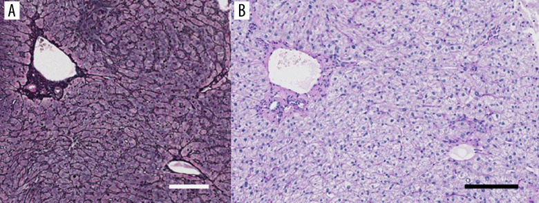
Figure 2.
The histological examination of the liver biopsy specimen. (A) The lobular architecture is well preserved. Grocott’s methenamine sliver impregnation. Bar: 100 µm. (B) Some lymphocytic infiltrations are seen in the sinusoids as well as the portal area, although no fibrous enlargement of the portal tract is seen. No inclusion bodies are found in the hepatocytes. Periodic acid-Schiff-diastase (PAS-D) reaction after diastase digestion. Bar: 100 µm.

