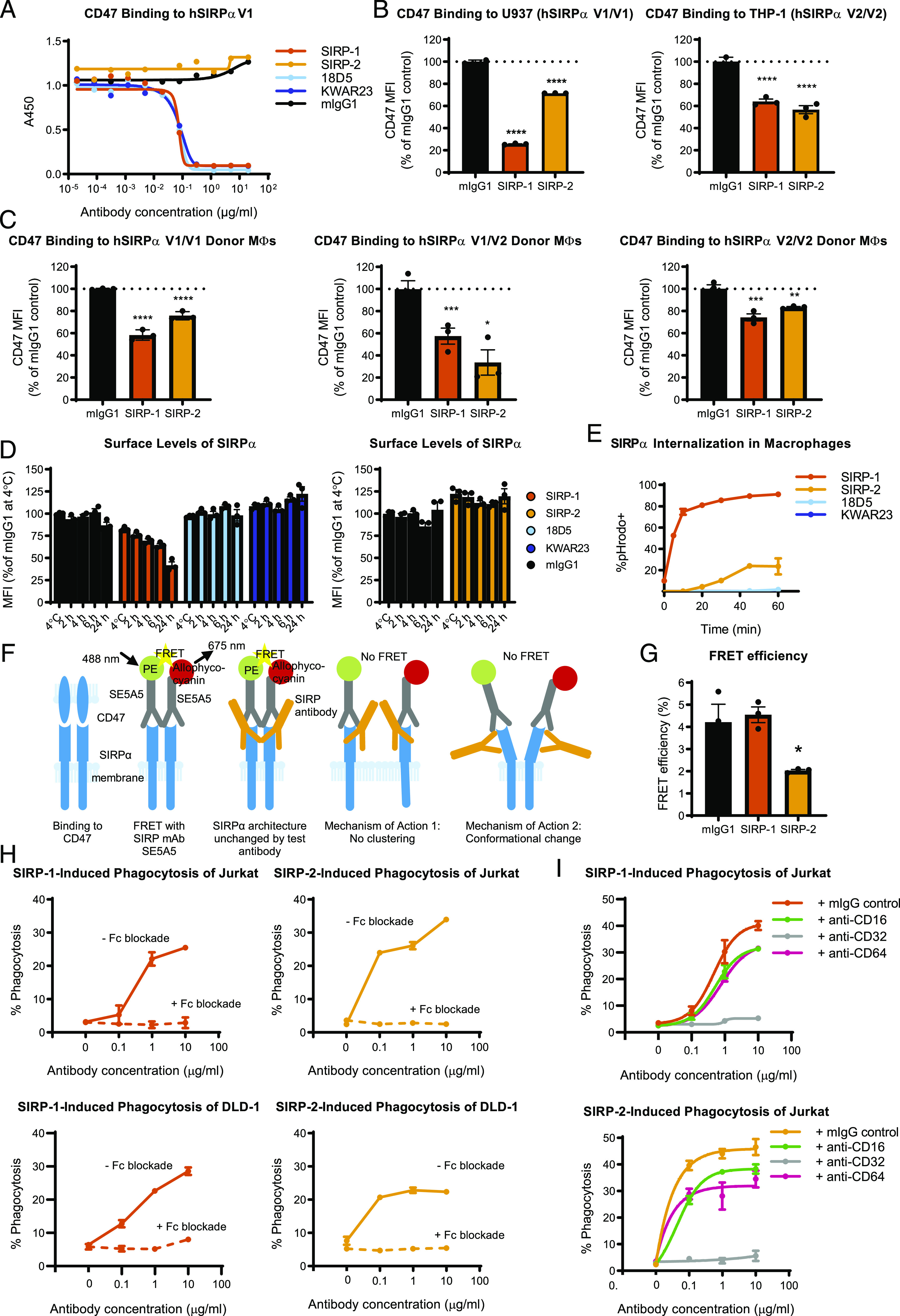
FIGURE 4.

SIRP-1 and SIRP-2 act via distinct mechanism of action: internalization and conformational change/declustering of SIRPα. (A) Blocking soluble CD47 binding to human SIRPαV1 (hSIRPαV1) was assessed by ELISA. Reduction of 20 μg/ml soluble CD47 binding to cell-expressed SIRPαV1 and SIRPαV2 by 10 μg/ml SIRP Abs was assessed using (B) promonocytic cell lines U937 and THP-1 and (C) moMΦs from various donor genotypes. (D) SIRP-1, but not mIgG1 control, 18D5, KWAR23, or SIRP-2 at 10 μg/ml reduced surface SIRPα levels in moMΦs in a time-dependent manner. The cells were incubated with Abs for 2 h at 4°C or 2, 4, 6, or 24 h at 37°C. (E) A total of 10 μg/ml SIRP-1 and, to a lesser extent, SIRP-2 caused Ab internalization in a time-dependent manner when measured by the uptake of pHrodo dye–Ab conjugates. (F) SIRPα binds CD47 most efficiently when clustered. When SIRPα Ab clone SE5A5, labeled with PE or allophycocyanin in equimolar concentrations, is added to cells, FRET is observed if SIRPα molecules and Abs bound to them are in close proximity. When a noncompeting anti-SIRP Ab is added that does not alter SIRPα architecture, no reduction in FRET is seen. No FRET is observed when a noncompeting anti-SIRP Ab that disables SIRPα from clustering is added. No FRET also occurs if binding of a noncompeting SIRPα mAb induces a conformation change in SIRPα that increases the distance between SE5A5 epitopes. (G) Treatment of moMΦs with 10 μg/ml SIRP-2, but not SIRP-1, for 2 h under the same conditions as in phagocytosis assays reduced the FRET efficiency between anti-SIRP Abs SE5A5-PE and SE5A5–allophycocyanin, indicating SIRPα conformational change/receptor declustering upon SIRP-2 treatment. (H) Addition of 10 μg/ml blocking Abs against human CD16, CD32, and CD64 inhibits SIRP-1– and SIRP-2–induced phagocytosis of Jurkat and DLD-1 cells by human moMΦs. (I) Function-blocking Abs against CD32 at 10 μg/ml, but not CD16 or CD64, inhibit SIRP-1– and SIRP-2–mediated Jurkat phagocytosis of moMΦs. All panels show mean ± SEM; a representative minimum n = 2 is shown. Statistical differences compared with the mIgG1 control are indicated. *p < 0.05, **p < 0.01, ***p < 0.01, ****p < 0.0001.
