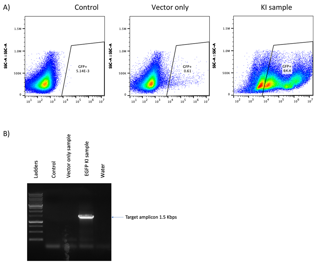
Figure 5. CRISPR/Cas9 and rAAV6 mediated site-specific integration of EGFP reporter cassette in B cells at day 12 post engineering.

(A) Representative flow plot shows no EGFP-positive B cells in either the control or the “vector only” sample and 64.4% in EGFP B cell was observed in the KI sample. (B) Junction PCR of KI samples shows 1.5 Kbps band which is the predicted size of the amplicon while no band was found in the control or “vector only” samples. A water lane is used to ensure no contamination in the PCR process.
