
FIGURE 7:
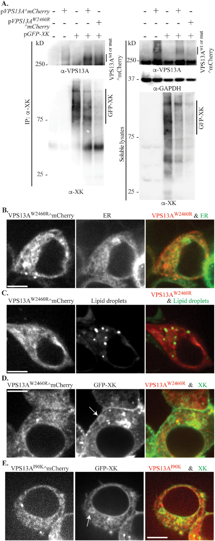
The disease mutation W2460R alters VPS13A^mCherry localization (A) Lysates of untransfected HEK293T cells, or cells transfected with single plasmids expressing VPS13A^mCherry (pVPS13A^mCherry) or GFP-XK (pcDNA3.1(+)-N-eGFP-XK), or two plasmids expressing wild-type VPS13A^mCherry or VPS13AW2460R^mCherry (pJS141-E4) with GFP-XK were analyzed by co-IP using polyclonal anti-XK antibodies and Western blot using anti-VPS13A antibodies, anti-XK antibodies, or anti-GAPDH antibodies as a loading control. (B) Localization of VPS13AW2460R^mCherry was examined in HEK293T cells cotransfected with plasmids expressing VPS13AW2460R^mCherry (pJS141-E4) and the ER marker mTagBFP-KDEL (pEFIRES-mTagBFP-KDEL). (C) VPS13AW2460R^mCherry localization was examined in HEK293T cells stained with the lipid droplet dye BODIPY 493/503. (D) Distribution of VPS13AW2460R^mCherry in cells cotransfected with plasmids expressing VPS13AW2460R^mCherry (pJS141-E4) and GFP-XK (pcDNA3.1(+)-N-eGFP-XK). (E) Distribution of VPS13AI90K^mCherry in cells cotransfected with VPS13AI90K^mCherry (pJS144-B2) and GFP-XK (pcDNA3.1(+)-N-eGFP-XK). Arrows in D and E indicate rings of GFP-XK in the ER. Scale bars = 10 μm.