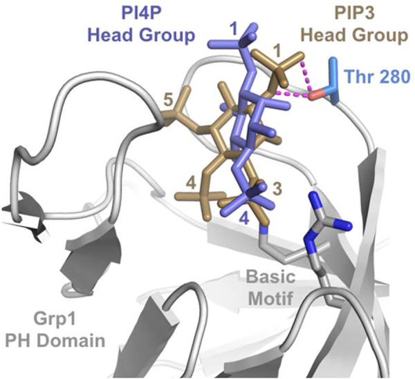
FIGURE 6:

Structural model for the differential effect of T280 phosphorylation on PIP3 vs. PI4P binding to the Grp1 PH domain. The phosphoinositide-binding site in the crystal structure of the Grp1 PH domain bound to the PIP3 head group (PDB ID 1FGY) is compared with a hypothetical composite model for the PI4P head group. The PI4P head group was acquired from the crystal structure of the ARNO PH domain bound to the PIP2 head group (PDB ID 1U29) after alignment of the PH domains and deletion of the 5-phosphate. Magenta dashes represent hydrogen bonds between the T280 side chain hydroxyl group and the 1-phosphate of the PIP3 head group that are not observed for the PIP2 head group in the ARNO complex.
