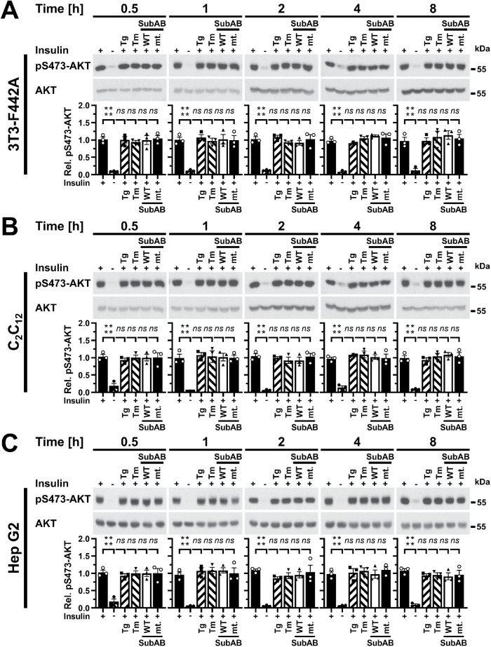
FIGURE 2:
Acute ER stress does not inhibit phosphorylation of AKT on S473 stimulated with 10 nM insulin for 15 min. (A) 3T3-F442A cells, (B) C2C12 myotubes, and (C) Hep G2 cells were serum-starved for 18 h and treated with 0.3 μM thapsigargin, 1 μg/ml tunicamycin, 1 μg/ml SubAB, or 1 μg/ml catalytically inactive SubAA272B during the last 30 min of serum starvation and then stimulated with 10 nM insulin for 15 min where indicated. Cell lysates were analyzed by Western blotting. Bars represent SEs (n = 3). p values for comparison of ER-stressed samples and samples not stimulated with 10 nM insulin to samples stimulated with 10 nM insulin were calculated by ordinary two-way ANOVA with Dunnett’s multiple comparisons test.

