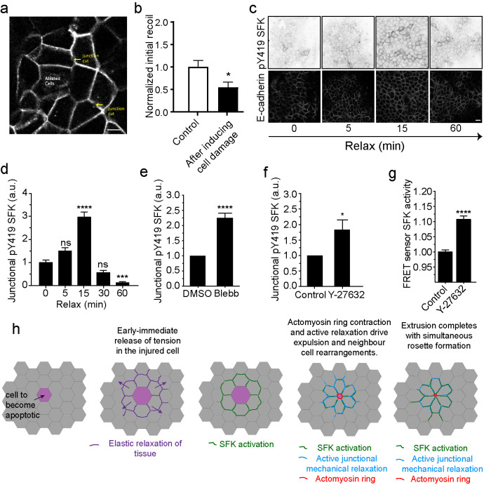
FIGURE 4:
SFK signaling is activated by elastic relaxation of neighboring cells. (a and b) Schematics of junctional tension measurements in the epithelium surrounding cells that were induced to cell death on ablation (ablated cells) (a). Initial recoil measurements were measured on cell junctions in either undamaged monolayers (Control) or in the neighborhood of ablated cells (after inducing cell damage). Data are mean ± SEM, for n = 20 initial recoil measurements per condition. *p < 0.05; unpaired t test. (c and d) pY419 SFK and E-cad-immunostained monolayers at different time points after mechanical relaxation of the underlying substrate (see Materials and Methods) and quantification of pY419 SFK at different time points Data are mean ± SEM, n = 100 junctions per condition; ***p < 0.001; ****p < 0.0001, one-way ANOVA, Tukey’s multiple comparisons test. Data representative of three independent experiments. (e and f) pY419 SFK junctional content in control or (e) blebbistatin (Blebb) or (f) Y-27632 (Y-27632)-treated monolayers. Data are mean ± SEM, n = 20 (e) and n = 8 (f) independent experiments; ****p < 0.0001, *p < 0.05, paired two-tailed t test. (g) Src activity (FRET index) in Src-Bio-tK MCF-7 cells treated (Y-27632) or not (Control) with Y-27632. Data are mean ± SEM, n = 4 independent experiments, at least 50 junctions per experiment were analyzed; ****p < 0.0001, paired two-tailed t test. Scale bars, 20 µm. (h) Model of SFK signaling activation in response to elastic relaxation of the tissue around apoptotic cells and how does it contribute to rosette formation and apoptotic cell extrusion.

