Figure 2:

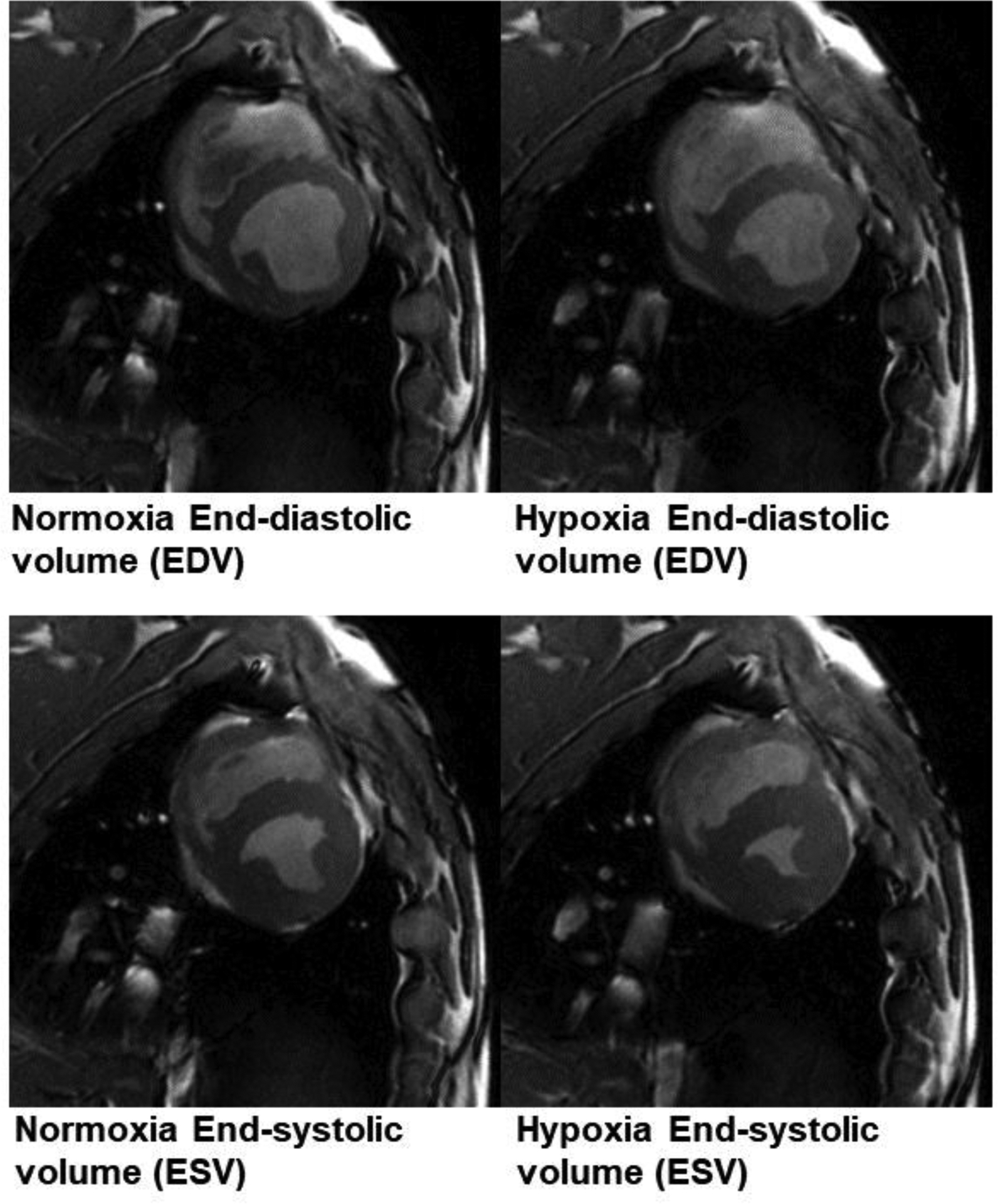
Example cine MR imaging at end diastole (top) and end systole (bottom). During hypoxia, there was a decline in both the end-diastolic volume (EDV) and end-systolic volume (ESV). ESV declined to a greater extent than did EDV.

Example cine MR imaging at end diastole (top) and end systole (bottom). During hypoxia, there was a decline in both the end-diastolic volume (EDV) and end-systolic volume (ESV). ESV declined to a greater extent than did EDV.