
Figure 7:
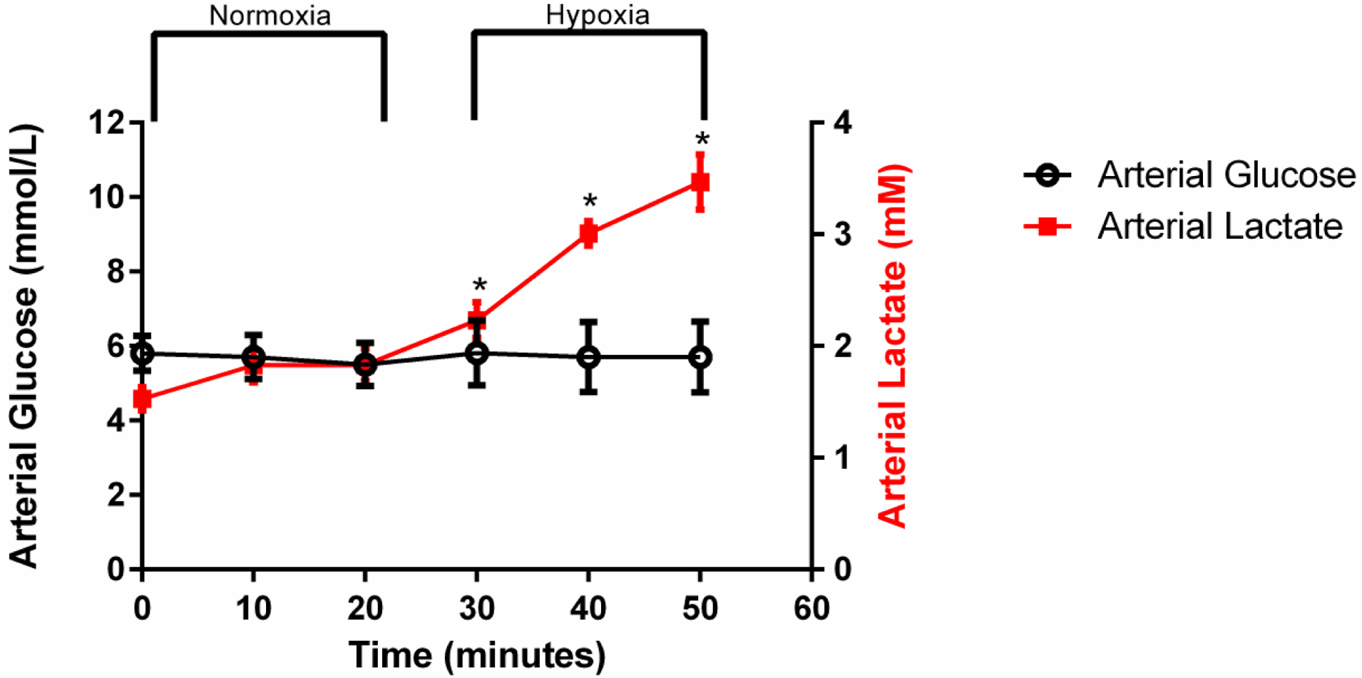
Plasma obtained from the carotid artery was used to determine changes in glucose and lactate during the rest-stress protocol. There were no differences in arterial plasma glucose over time, while hypoxia induced an increase in arterial plasma lactate. Values represent Mean ± SEM. * indicates significant increases in arterial plasma lactate during hypoxic stress compared to resting conditions (p < 0.05)