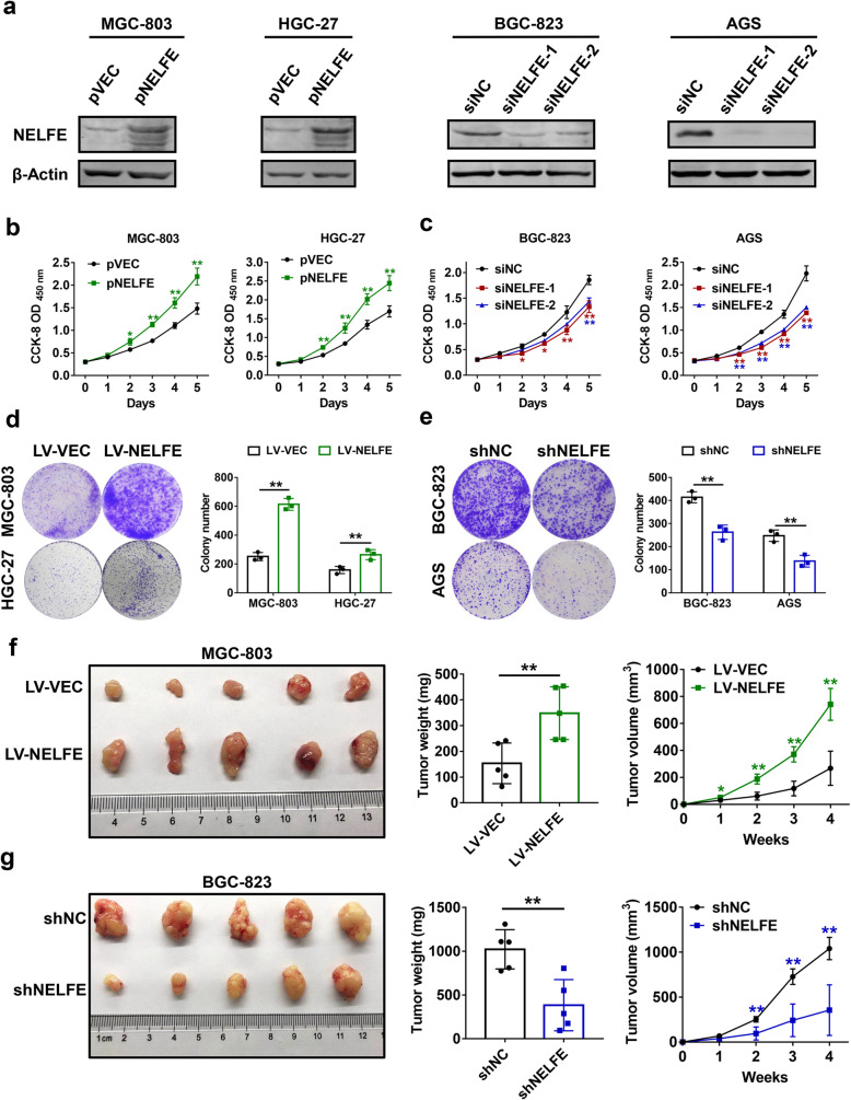
Fig. 2.
NELFE potentiates GC cell proliferation in vitro and in vivo. a NELFE-expressing plasmids or siRNAs against NELFE were transfected into the indicated GC cell lines, respectively. After 48 h, cells were lysed and western blot analyses were performed to verify the efficacies of overexpression and knockdown. b-c Cell viabilities were determined using CCK-8 cell proliferation assays at every 24 h after transfection of the above plasmids (b) or siRNAs (c). d-e Colony formation assays were conducted using the indicated GC cell lines with lentivirus-mediated stable overexpression (d) or knockdown (e) of NELFE. Representative colony images (left) and statistical analysis of the colony number in different groups (right) were shown. f-g Subcutaneous xenograf model of nude mice was established using MGC-803 or BGC-823 cells with stable overexpression (f) or knockdown (g) of NELFE (n=5 per group). Images of tumors (left), final tumor weight (middle) and tumor volume curve (right) were shown. *P < 0.05, **P < 0.01

