FIGURE 8:
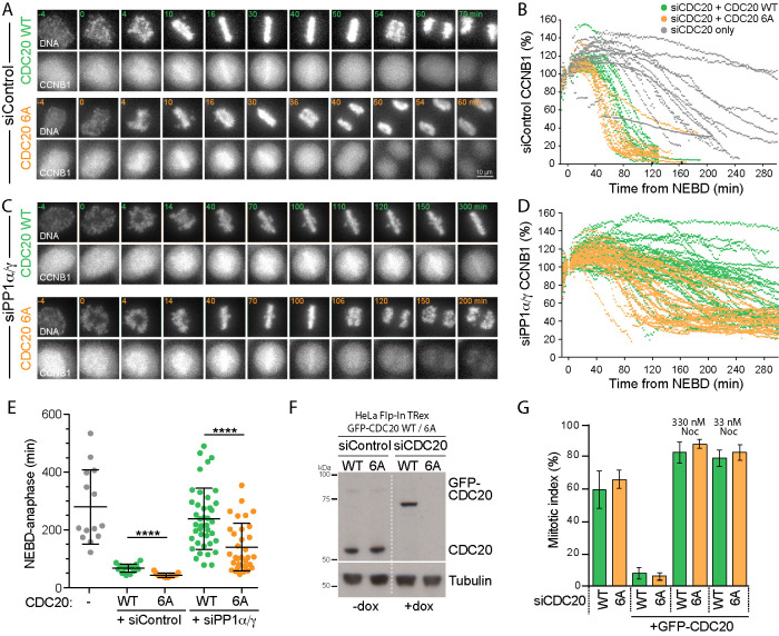
CDK1 phosphorylation site mutant CDC206A bypasses the requirement for PP1 in rapid cyclin B destruction on mitotic exit. (A) HeLa Flp-In TRex cells expressing CCNB1-mCherry were depleted with siControl and siCDC20 and then induced to express either GFP-CDC20WT or GFP-CDC206A. Cell cycle progression and CCNB1-mCherry levels were followed by live cell imaging. DNA was visualized with SiR-DNA. (B) CCNB1 levels for individual cells are plotted in the line graph. Gray lines show CCNB1 levels in cells depleted with siCDC20 but without induction of the GFP-CDC20 transgene. (C) HeLa Flp-In TRex cells expressing CCNB1-mCherry were codepleted of siPP1α/γ and siCDC20 and then induced to express either GFP-CDC20WT or GFP-CDC206A. Cell cycle progression and CCNB1-mCherry levels were then followed by live cell imaging. DNA was visualized with SiR-DNA. (D) CCNB1 levels for individual cells are plotted in the line graph. (E) Scatter plots showing the mean time ± SD at which cells entered anaphase or the end of the movie was reached for siCDC20 uninduced (n = 12, gray), siControl with GFP-CDC20WT (n = 20, green) or GFP-CDC206A (n = 15, orange), and siPP1α/γ with GFP-CDC20WT (n = 41, green) or GFP-CDC206A (n = 33, orange). (F) Western blot of cells depleted of endogenous CDC20 and expressing GFP-CDC20 as in A, arrested with 330 nM nocodazole for 14 h. **** denotes p < 0.0001. (G) Cells depleted for endogenous CDC20 (siCDC20) and induced for GFP-CDC20WT (WT) or CDC206A (6A) were treated with nocodazole at the indicated concentrations for 14 h, and the mitotic index was plotted.

