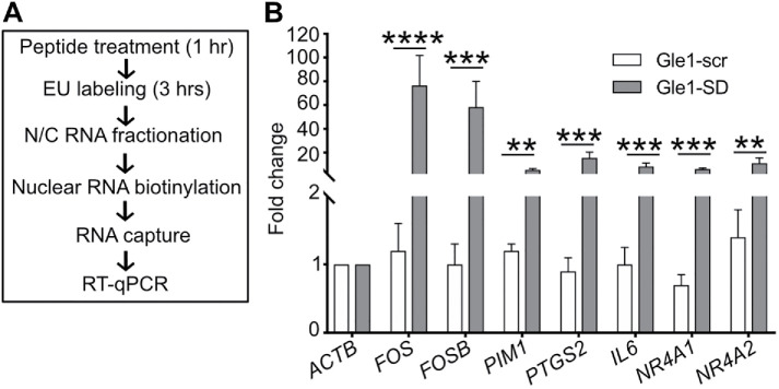
FIGURE 3:
Gle1-SD peptide treatment leads to accumulation of nascent mRNAs. (A) Schematic diagram illustrates EU-based detection of nascent mRNA. (B) HeLa cells were labeled with EU in the presence of Gle1-scr or Gle1-SD peptide. Nascent nuclear RNAs were captured and subjected to RT-qPCR using primers in the CDS region. The graph depicts fold change relative to actin values in untreated samples (mean ± SEM) from three biological replicates performed in triplicate (as detailed in Figure 1). ΔCT values were used to calculate statistical significance using paired one-tailed Student t test (****p < 0.0001, ***p < 0.001, **p < 0.003).

