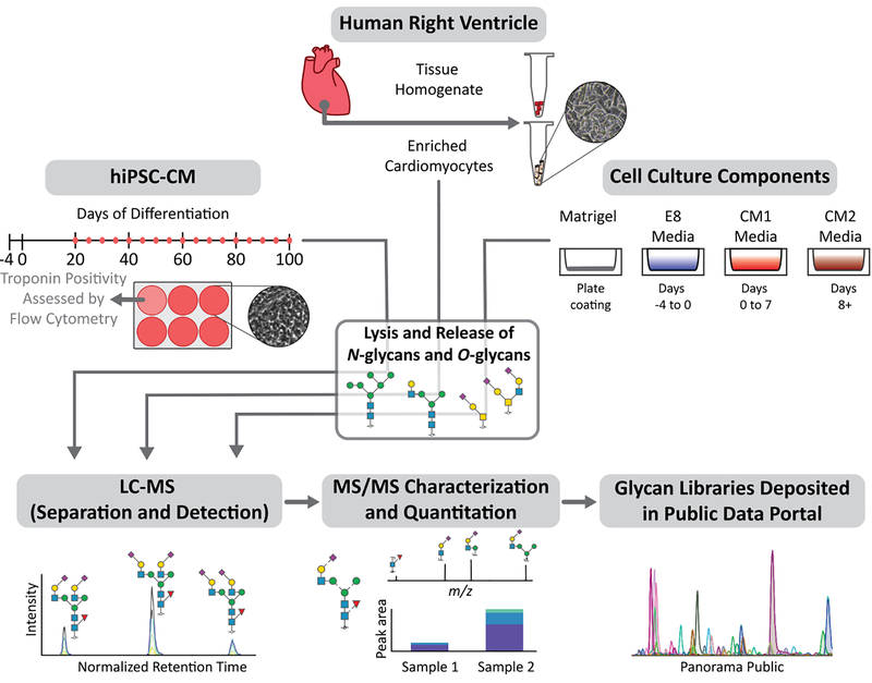
Figure 1.
Schematic outline of the study design investigating protein glycosylation in human cardiomyocytes. Glycans from cardiomyocytes enriched from human heart tissue (n=3), matched tissue homogenate (n=3), hiPSC-CM collected across days 20–100 of differentiation (n=3 for each time-point), and hiPSC-CM culture components (n=1) were analyzed using PGC-LC-ESI-MS/MS for structure-based glycan characterization and quantitation. Glycan structure libraries are publicly available in Panorama to facilitate future glycoproteomic and glycomic efforts.

