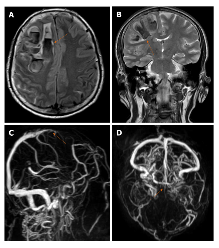
Figure 2.
A 61-year-old male with Dural venous thrombosis. A and B: Axial and coronal sections of magnetic resonance imaging brain (T2WI sequence) show acute hemorrhage (arrow) in right frontal lobe with left sided midline shift; C and D: Magnetic resonance venography sagittal oblique and axial sections show absent flow related signal in anterior third of superior sagittal sinus suggestive of thrombosis.

