
Fig. 2. PA/KN hydrogel combinations and material analysis.
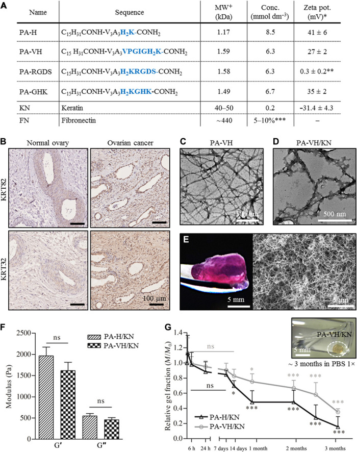
(A) Table of PA sequences and proteins used in this study including the chemical formula, molecular weight (MW), concentration used, and zeta potential at pH 7.5 (*in HEPES (pH 7.5), **in water, ***relative to keratin volume, +theoretical). (B) Immunohistochemical (IHC) staining for keratin type I (KRT32) and type II (KRT82) in normal (healthy) ovary and ovarian cancer tissue samples, with IHC controls in fig. S2. (C) TEM image of self-assembled PA fibers (PA-VH) in HEPES [0.1 mg/ml (pH 7.5)]. (D) TEM image of PA fibers interacting with KN (PA-VH/KN). Pure solutions of PA-VH and KN in HEPES [0.1 mg/ml (pH 7.5)] were mixed in ratio 1:1 before analysis. (E) Photograph of a PA-H/KN hydrogel after 14 days in culture medium at 37°C and a representative scanning electron microscopy image of the internal heterogeneous nanofibrous structure of PA/KN hydrogels. (F) Rheological characterization of the hydrogels, measuring the storage modulus (G′) and loss modulus (G″) of PA-H/KN and PA-VH/KN hydrogels (mean ± SEM). (G) Graphical plot of the hydrogel mass (Mt/M0) against time, indicating the slow degradation of PA-H/KN and PA-VH/KN hydrogels in PBS 1× at 37°C (mean ± SEM). Photo credit: Clara Hedegaard, Queen Mary University of London. ns, not significant.