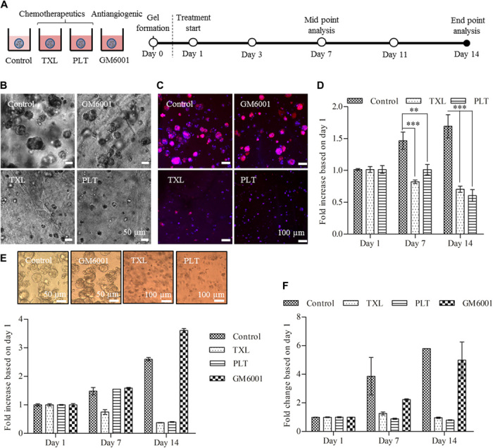
Fig. 6. Treatment study with TXL, PLT, and GM6001 using cells grown in PA-VH/KN hydrogels and Matrigel.

(A) Schematic showing the experimental setup for the treatment studies. Ovarian cancer cells (OVCAR-4) were encapsulated at a density of 500 cells/μl with treatment starting 1 day after encapsulation. (B) Bright-field images of the tumor spheroid sizes after 14 days for the control and three treatments. (C) IF images of the F-actin network (phalloidin/red) and nuclei (DAPI/blue) of tumor spheroids on day 14 for all treatment conditions. The IF images correspond to the bright-field images in (B). (D) Metabolic activity of cells grown in PA-VH/KN hydrogels for all treatments over 14 days (values normalized to day 1), measured using an almarBlue assay. (E) Metabolic activity of cells grown in Matrigel for all treatments over 14 days (values normalized to day 1), measured using an almarBlue assay. (F) Metabolic activity of tricultures (ovarian cancer cell/HUVEC/hMSC ratio of 560:6000:600) in PA/KN hydrogels for all treatments over 14 days (values normalized to day 1), measured using almarBlue assay.
