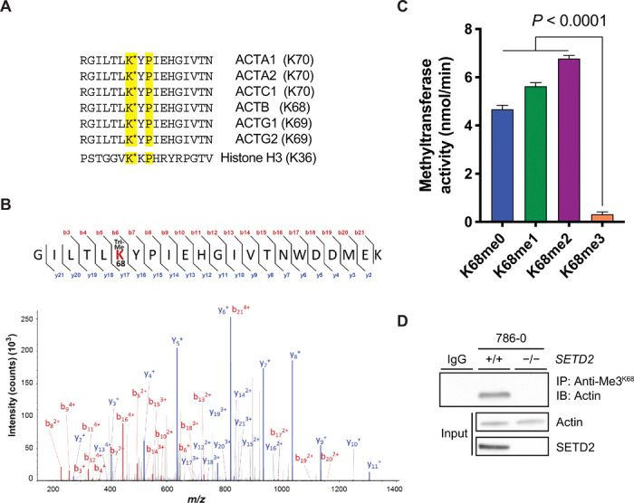
Fig. 3. SETD2 methylates lysine-68 on actin.

(A) Amino acid sequence showing conserved KxP SETD2 recognition motif present in all actin isoforms. Position of the lysine residue varies depending on actin isoform; reference to this site as “ActK68” is based on its position in β-actin (ACTB). Histone H3 sequence containing the KxP motif is shown below for reference. (B) Representative tandem mass spectrometry (MS/MS) spectrum of trimethylated ActK68 peptides recovered from SETD2-proficient 786-0 cells. m/z, mass/charge ratio. (C) Fluorescence-based in vitro methylation assay showing in vitro methylation of biotin-labeled K68-containing actin peptides (amino acids 62 to 78) with recombinant tSETD2 (amino acids 1418 to 2564). Sequence for the peptides used is shown in (A). Data are means ± SEM (n = 4). (D) Immunoblot analysis showing dependency of the ActK68me3 mark on SETD2 by IP of endogenous actin from whole-cell extracts of SETD2-proficient and SETD2-deficient 786-0 cells using the anti-Me3K68 antibody. Data are representative of experiments repeated at least three times with similar results.
