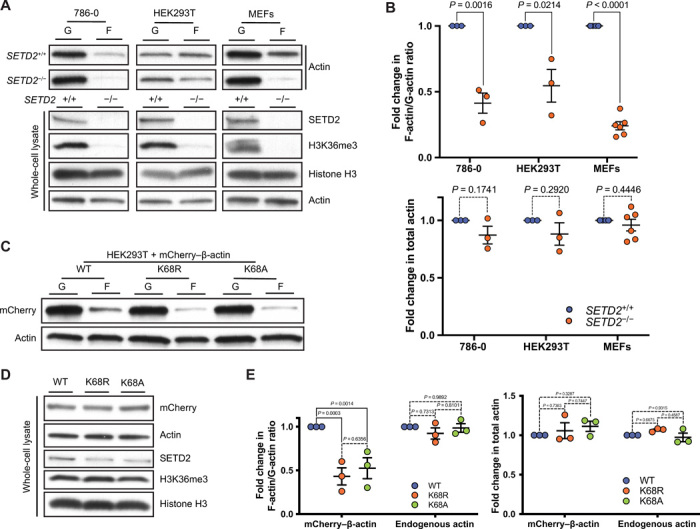
Fig. 4. SETD2 regulates actin polymerization in cells.

(A) Immunoblot analysis showing decreased F-actin in SETD2-deficient 786-0, HEK293T, and MEF cells. Whole-cell lysate shows absence of SETD2, associated with the expected loss of histone H3K36me3 methylation. (B) Quantitation of F-/G- actin ratio (top) and whole-cell lysate actin (bottom) from the data is shown in (A). Data are means ± SEM (n = 3 for 786-0 and HEK293T; n = 6 for MEF). (C) Immunoblot analysis of F-/G-actin ratio in HEK293T cells expressing wild-type or K68A/R mCherry–β-actin. (D) Immunoblot analysis of whole-cell lysates shows no change in total actin levels or SETD2 histone methylation with expression of K68A/R mCherry–β-actin. (E) Quantitation of F-/G-actin and total actin shown in (C) and (D), respectively. Data are means ± SEM (n = 3).
