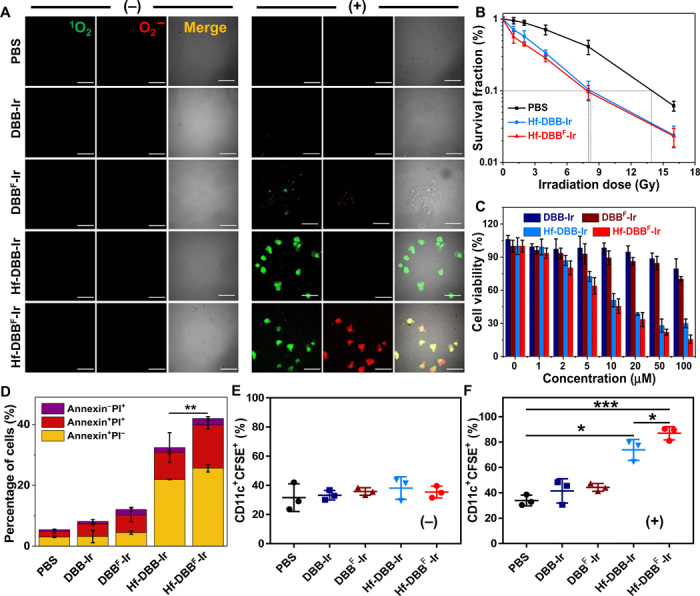
Fig. 2. In vitro generation of DAMPs and phagocytosis.

(A) Generation of 1O2 and O2− in MC38 cells treated with phosphate-buffered saline (PBS), DBB-Ir, DBBF-Ir, Hf-DBB-Ir, or Hf-DBBF-Ir with (+) or without (−) x-ray irradiation as detected by SOSG and superoxide kits. Green (1O2) and red (O2−) florescence merged to appear as yellow florescence. Scale bars, 50 μm. (B) Clonogenic assays to evaluate radioenhancement of Hf-DBB-Ir or Hf-DBBF-Ir on MC38 cells upon x-ray irradiation; n = 6. (C) Cytotoxicity of Hf-DBB-Ir or Hf-DBBF-Ir upon x-ray irradiation at a dose of 2 Gy on MC38 cells; n = 6. (D) Annexin V/propidium iodide (PI) cell apoptosis/death analysis of MC38 cells; n = 3. (E and F) Phagocytosis of carboxyfluorescein diacetate succinimidyl ester (CFSE)–labeled MC38 cells by DCs. DCs cocultured with treated MC38 cells without (−) (E) or with (+) (F) irradiation were stained with PE-Cy5.5–conjugated CD11c antibody; n = 3. Data are expressed as means ± SD. *P < 0.05, **P < 0.01, and ***P < 0.001 by t test.
