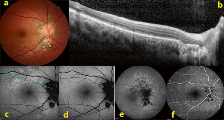
Fig. 1. Fundus autofluorescence in a case of multifocal choroiditis relapse.
a Baseline colour ophthalmoscopic image showing chorioretinal scars around the optic disc. b SD-OCT with disruption of the ellipsoid zone through the bright autofluorescent area. c BAF imaging showing increased autofluorescence around the optic disc and above the macula. d This hyperautofluorescence disappeared after photobleaching process. e ICGA late frame showing confluent dark hypofluorescent area around the optic disc and numerous hypofluorescent dots representing areas of new inflammatory involvement. The optic disc and the chorioretinal scars were black. f FA showing only small hypofluorescent areas under the optic disc corresponding to chorioretinal scars.

