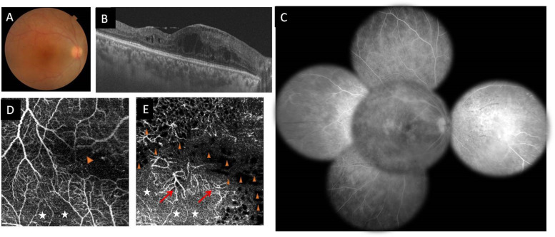
Fig. 12. Multimodal imaging of the right eye of a 32-year-old female patient with Behçet disease.
a Colour fundus photography showing vascular sheathing; b swept source OCT B-scan showing macular oedema with cystoid spaces. c Composite fundus fluorescein angiogram of the same eye showing diffuse fern-like capillary leakage with associated peripheral nonperfused areas nasally and temporally. d 3 × 3 mm swept source OCT angiography scan of the superficial capillary plexus clearly visualizes well-delineated hypointense greyish areas of retinal capillary nonperfusion (white asterisk) and perifoveal capillary arcade disruption (arrowhead). e Swept source OCT angiogram shows in the deep capillary plexus, larger greyish areas of capillary nonperfusion (white asterisk), well-defined black roundish areas completely devoid of flow (arrowheads), and capillary telangiectasias (red arrows).

