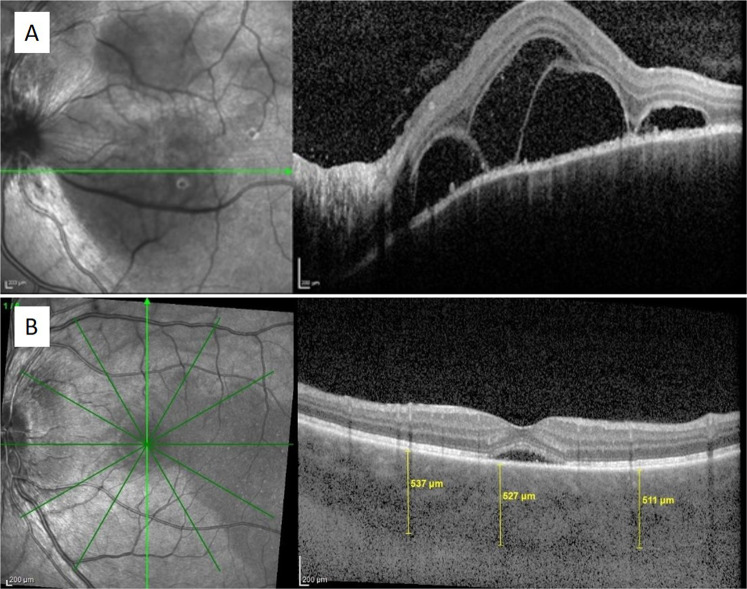
Fig. 9. Retinal and choroidal OCT features in acute Vogt-Koyanagi-Harada disease.
SD-OCT shows serous retinal detachment with subretinal septae in the left eye of a patient with acute Vogt–Koyanagi–Harada disease (a). EDI-OCT in another acute case of Vogt–Koyanagi–Harada disease shows substantial thickening of the choroid 511–537 µm (b).

