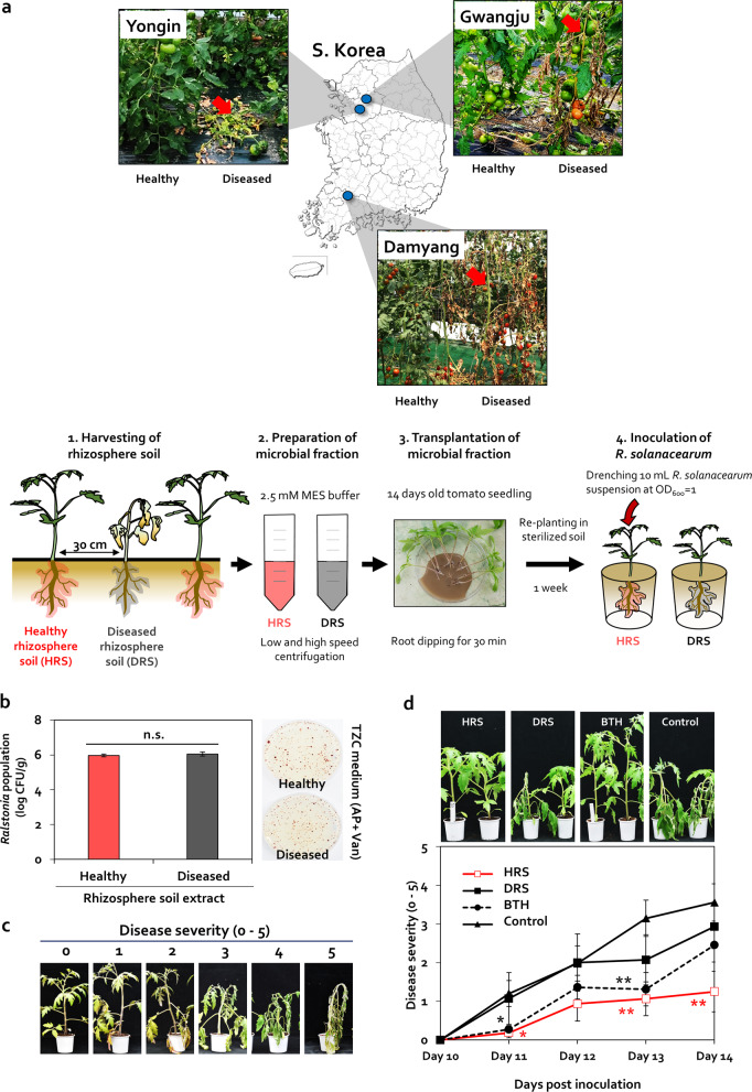
Fig. 1. Differences in rhizosphere disease suppression between two adjacent tomato plants.
a Images of healthy and diseased tomato plants grown within a 30-cm distance in three different locations (Damyang, Yongin, and Gwangju) in South Korea. Red arrows indicate the wilted tomato plants infected by Ralstonia solanacearum. To prepare the microbial fraction, healthy rhizosphere soil (HRS) and diseased rhizosphere soil (DRS) were suspended in 2.5-mM MES buffer. Roots of 14-day-old tomato seedlings were dipped in the microbial fractions for 30 min. Severity of bacterial wilt disease caused by R. solanacearum was quantified. Data represent mean ± standard error of the mean (SEM; n = 12 plants per treatment). Asterisks indicate significant differences (*P < 0.05, **P < 0.01, ***P < 0.001). b Cell density of R. solanacearum in HRS and DRS fractions plated on casamino acid-peptone-glucose (CPG) agar medium containing 2,3,5-triphenyl tetrazolium chloride (TZC), 0 or 50-g/mL ampicillin (AP), and 0 or 5-g/mL vancomycin (Van). c Scoring of disease severity on a 0–5 scale. d Disease severity in HRS and DRS fraction-treated tomato plants at 10–14 days post inoculation (dpi). BTH, 0.5-mM benzothiadiazole treated tomato; control, 2.5-mM MES buffer-treated tomato.

