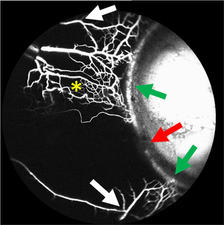
Fig. 1. Segmental aqueous humour outflow.

ICG aqueous angiography was performed in an 82-year-old Caucasian man’s left eye. The aqueous humour outflow pattern demonstrated regions of high-flow (green arrows) and low-flow (red arrow). Y-shaped episcleral and aqueous veins were seen (white-arrows) in addition to a network of vessels likely representing the intrascleral venous plexus (yellow asterisk) (color figure online).
