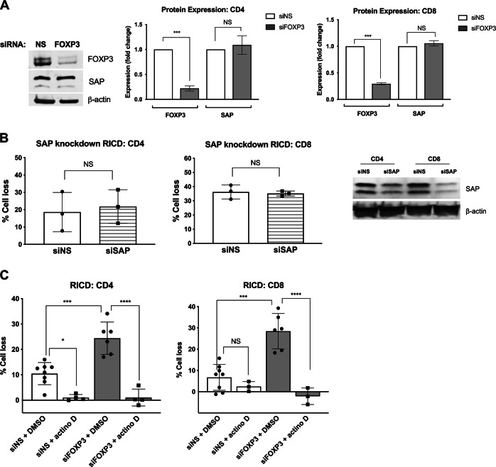
Fig. 2.
RICD in expanding T cells is SAP-independent, but requires de novo transcription. a Day 4 FOXP3-KD cells were analyzed for FOXP3 and SAP expression by western blotting (left panel). Protein expression was quantified after normalization to β-actin (right panels). The data represent three independent experiments. Statistical significance was determined by one-way ANOVA with Sidak’s multiple comparisons test. CD4: ***p < 0.001, NS not significant. CD8: ***p < 0.001, NS not significant. b Purified CD4 and CD8 T cells were electroporated with SAP-specific siRNA or a nonspecific (NS) control. Activated T cells were restimulated on day 4 with 100 ng/mL OKT3, and RICD was assessed by PI staining and flow cytometry. Gene knockdown was verified by western blotting (right). Statistical significance was assessed with paired t tests, NS not significant. c Day 4 KD T cells were pretreated with 100 ng/mL actinomycin D (actino D) or a DMSO solvent control for 30 min before restimulation with 100 ng/mL OKT3. Statistical significance was determined by one-way ANOVA with Sidak’s multiple comparisons test. CD4: *p = 0.0198, ***p = 0.001, ****p < 0.0001. CD8: ***p = 0.001, ****p < 0.0001

