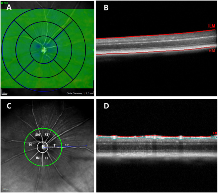
Figure 1.
(A) Concentric rings with 1, 2, and 3 mm diameters. (B) OCT section. Retinal thickness measured between the inner limiting membrane and the retinal pigment epithelium. (C) RNFL sectors (RNFL, Retinal Nerve Fiber Layer; ST, Supero–Temporal; SN, Supero–Nasal; N, Nasal; IN, Infero–Nasal; IT, Infero-Temporal; T, Temporal; G, Global). (D) OCT section. Result of segmentation of the OCT scan. RNFL delimited between the ILM and GCL + IPL. OCT, optical coherence tomography; RNFL, Retinal nerve fiber layer; ILM, inner limiting membrane; GCL + IPL, ganglion cell layer and inner plexiform layer.

