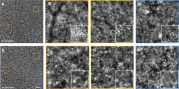
Fig. 8.
Photoreceptor imaging in an age-matched normal subject (a–d) and a retinitis pigmentosa patient (e–h). a, e Central 10° AO image. Orange, yellow and blue squares correspond to the magnified images in (b, f), (c, g) and (d, h), respectively. Decreased cone density and increased spacing can be detected in the patient. Reprinted with permission from Lin et al. [154].

