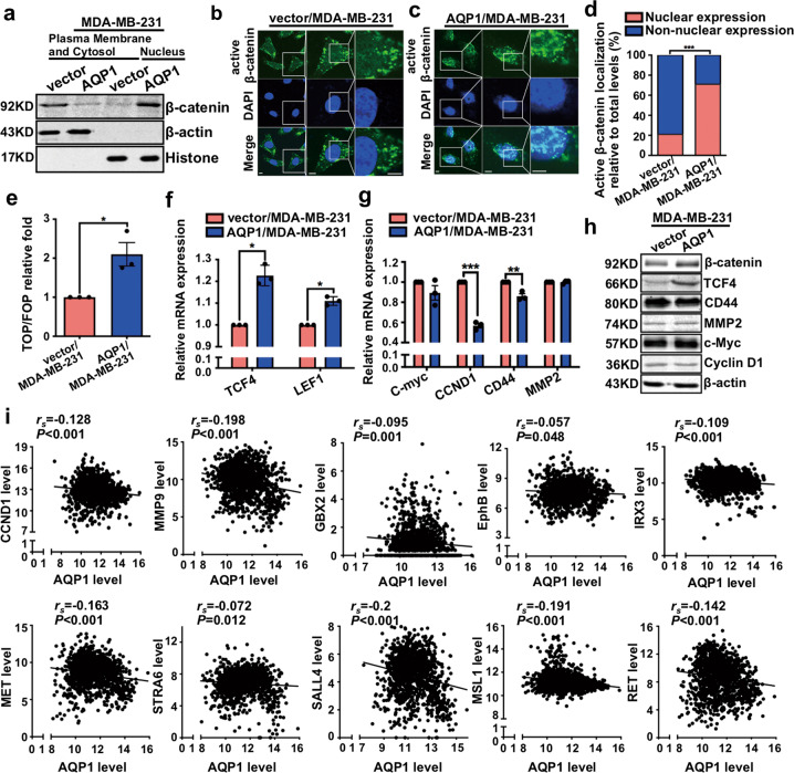
Fig. 5. AQP1-induced β-catenin nuclear translocation did not activate Wnt downstream target genes.
a Western blot analysis of β-catenin expression in plasma membrane, cytosol, and nucleus of breast cancer cells. β-actin and histone were used as specific markers for cytoplasm and nuclei, respectively. b, c Immunofluorescence analysis showed the localization of active β-catenin in vector/MDA-MB-231 (b) and AQP1/MDA-MB-231 (c) cells. Insets showed a high-magnification view of the indicated region. Scale bars: 50 μm. d Ratio of nuclear expression of active β-catenin was shown in a graph representation (χ2 test, ***P < 0.001). e Overexpression of AQP1 activated TOP-Flash reporter by TOP/FOP Flash experiments. Values were expressed as mean ± SEM (two-tailed Student’s t test, *P < 0.05). f Relative mRNA levels of TCF4 and LEF1 were quantified by RT-qPCR. Values were expressed as mean ± SEM (two-tailed Student’s t test, *P < 0.05). g Relative mRNA levels of C-myc, CCND1, CD44, and MMP2 were quantified by RT-qPCR. Values were expressed as mean ± SEM (two-tailed Student’s t test, **P < 0.01, ***P < 0.001). h Western blot for the indicated proteins were performed. β-actin was the loading control. i The 1218 breast cancer patients’ RNAseq data in TCGA was analyzed. AQP1 was negatively associated with Wnt downstream target genes, such as CCND1, MMP9, GBX2, EphB, IRX3, MET, STRA6, SALL4, MSL1, and RET. P value was calculated by Spearman’s Rank-Correlation test. Experiments a–h were independently repeated for three times.

