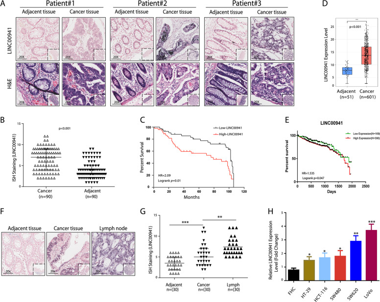
Fig. 1. LINC00941 expression is significantly upregulated during CRC development.
a Representative images of ISH staining of LINC00941 in CRC tissues and paired nontumor tissues of three CRC patients (n = 90). b The level of LINC00941 expression was calculated from CRC tissue arrays analyzed by ISH. c Kaplan–Meier curve depicting the overall survival of 90 CRC patients. d LINC00941 level in 601 CRC tumor samples and 51 normal controls from TCGA data. e Kaplan–Meier curve depicting the overall survival of 559 CRC patients from TCGA database. f and g Representative images of ISH staining of LINC00941 in adjacent normal tissues, tumor tissues, and positive metastatic lymph from CRC tissue arrays analyzed by ISH (n = 30). h The expression of LINC00941 in HCC cell lines was upregulated compared with that in the FHC immortalized human colorectal cell line, as shown by RT-qPCR. Data are presented as the mean ± SD. *p < 0.05, **p < 0.01, ***p < 0.001.

