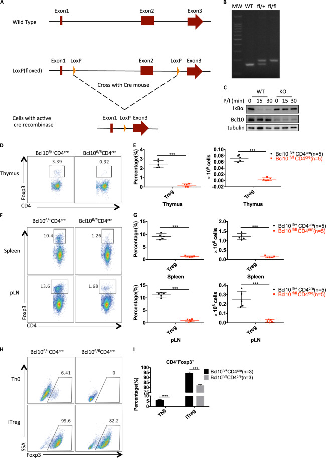
Fig. 1.
Bcl10 is required for the development of Treg cells. a The details are described in the methods and materials. A schematic graph of Bcl10fl/fl mouse generation is shown. Two loxP elements were inserted into introns 1 and 2. b Genotyping of WT (Bcl10+/+), fl/+ (Bcl10fl/+) and fl/fl (Bcl10fl/fl) mice was performed by PCR. c WT (from CD4cre/Bcl10+/+ mice) and Bcl10-conditional KO (from CD4cre/Bcl10fl/fl mice) pan T cells were stimulated with PMA (20 ng/mL) and ionomycin (200 ng/mL) for the indicated times, followed by immunoblotting analysis. d–g The thymus, spleen, and peripheral lymph nodes of 8- to 10-week-old CD4cre/Bcl10+/fl and CD4cre/Bcl10fl/fl mice were analyzed by FACS (n = 5 per group). d FACS plots show CD4+Foxp3+ cells gated on CD4+CD8− cells in the thymus. e Statistical analyses were used to evaluate the CD4+Foxp3+ cell percentage (left) in the CD4+CD8− cell population and absolute numbers (right) in the thymus. f FACS plots show CD4+Foxp3+ cells gated on CD4+CD8− cells in the spleen or peripheral lymph nodes. g Statistical analyses were used to evaluate the CD4+Foxp3+ cell percentage (left) in the CD4+CD8− cell population and absolute numbers (right) in the spleen or peripheral lymph nodes. h, i CD4+CD25−CD44−CD62L+ naive T cells sorted from a pooled spleen and lymph node suspensions from Bcl10+/flCD4cre or Bcl10fl/flCD4cre mice were stimulated by plate-bound anti-CD3/28 antibodies (5 μg/mL) alone (Th0) or in the presence of mouse IL-2 (10 μg/mL) and human TGF-β (2 ng/mL) (iTreg) for 3 days. Foxp3 induction in viable cells was detected in gated CD4+ cells h, and a summary of the Foxp3+ percentage is shown in i. Student’s t test was used as the statistical test (*p < 0.05, **p < 0.01, and ***p < 0.005). All error bars represent SDs

