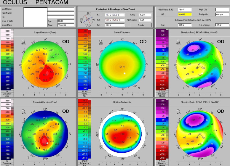
Fig. 3. Oculus Pentacam report of a patient with keratoconus.
Axial power and tangential maps are displayed in the left column, pachymetry and relative pachymetry map in the middle, and elevation of the anterior and posterior cornea relative to reference surfaces in the right column, with similar inferocentral “hot spot” regions across various maps indicative of keratoconus.

