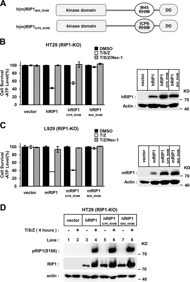
Fig. 4. RIP1M45_RHIM blocks necroptosis signal transduction downstream of RIP1 kinase activation.
a Schematic representation of chimaeric RIP1-containing M45/ICP6-RHIM. b, c RIP1 chimeras with M45–RHIM could not restore the sensitivity to necrotic stimuli in RIP1 knockout cells. RIP1 knockout HT-29 (b) or L929 (c) cells with indicated lentivirus infection were treated with indicated stimuli for 10 h (b) or 4 h (c). The final concentrations of 10 μm RIP1 inhibitor necrostatin-1 (Nec-1) were used to block necrosis. The number of surviving cells was determined by measuring ATP levels (left). The data are presented as the mean ± SD of duplicate wells. The RIP1 expression level was measured by western blot analysis (right). d TNF-induced RIP1 kinase activation is not affected in chimaeric RIP1. RIP1-KO HT-29 cells with indicated lentivirus infection were treated with the indicated stimuli for 4 h. The cells were harvested and analyzed by western blotting using antibodies as indicated.

