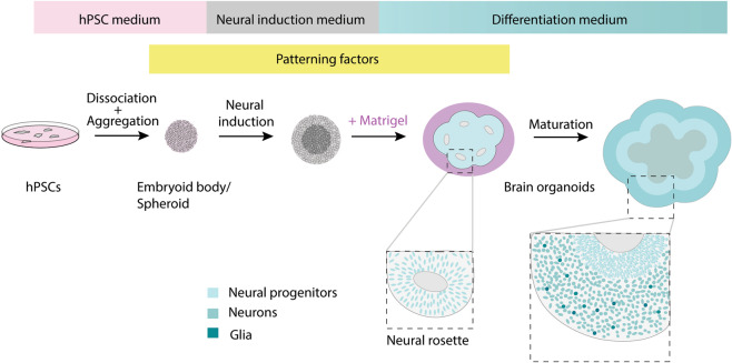
Fig. 1. Schematic representation of general brain organoid culture scheme.
Two-dimensional cultured hPSCs are dissociated and reaggregated to form an embryoid body (EB) or a spheroid in hPSC culture medium supplemented with Rho kinase inhibitor. Subsequent culture in a neural induction medium boosts the neuroectodermal fate. The neuroectodermal cells self-organize into multiple 3-D structures featuring apical lumens called “neural rosettes” or “neural buds,” reminiscent of the neural tube. This process is further boosted upon exposure to Matrigel, an extracellular matrix rich gel. Subsequently, the neuroectodermal cells give rise to the neural stem and progenitor cells that proliferate and differentiate to give rise to neurons and glia. The insets show zoomed in pictures with nuclei represented in shades of blue. For region-specific organoid protocols, patterning factors are added to the culture media at specific stages.

