
Figure 2. Crystal structure of compound 1 bound to E. coli PheRS explains structure-activity relationships.
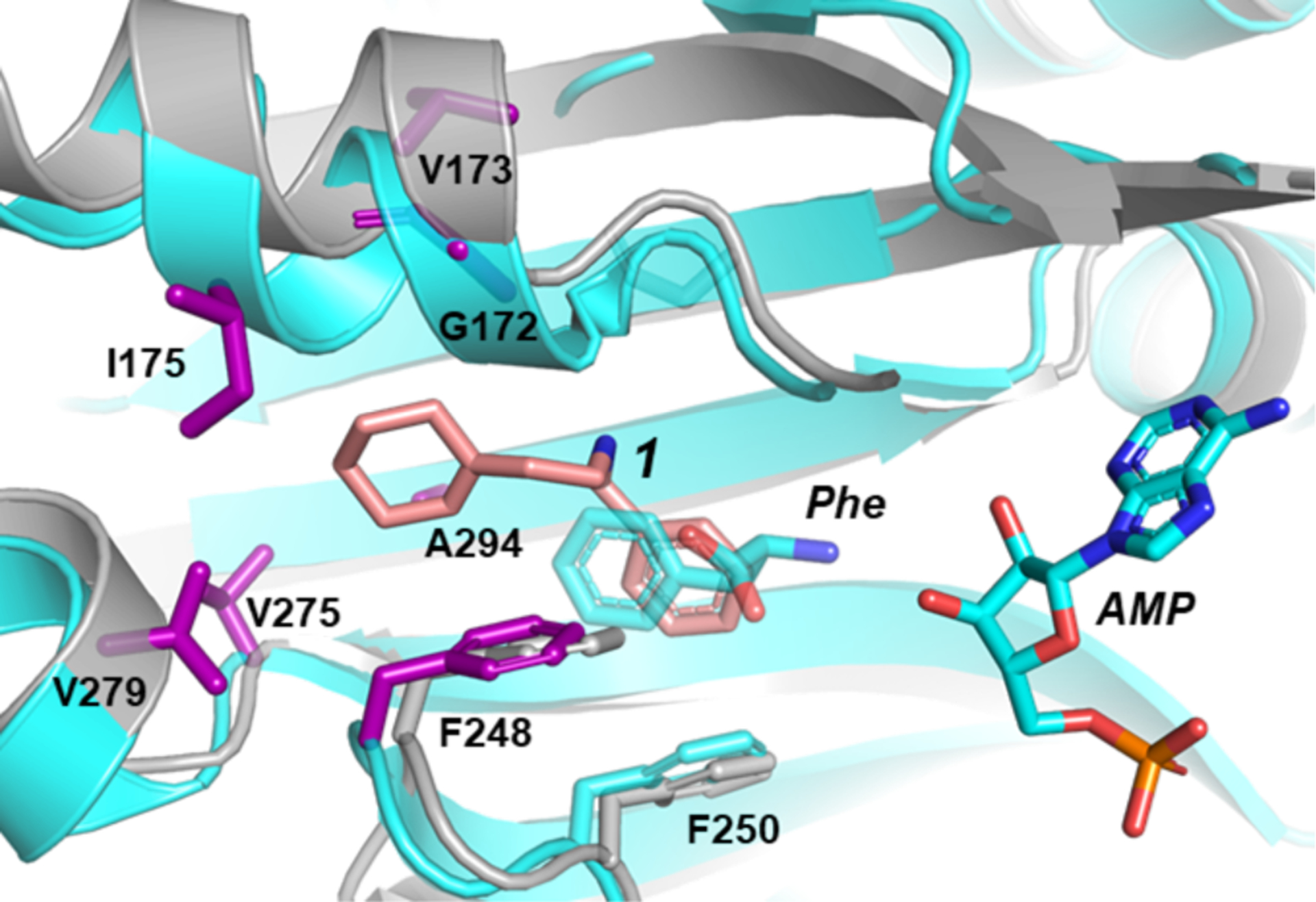
Cartoon and stick representations of two E. coli PheRS structures are overlain. The crystal structure of E. coli PheRS bound to phenylalanine and AMP (PDB #3PCO7) is colored cyan with residues at sites of resistance mutations shown as purple sticks. The structure of E. coli PheRS bound to compound 1 (salmon), is colored grey.