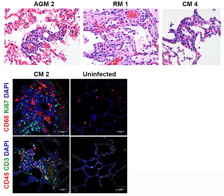
Fig 5. Histopathology and IFA.
Necropsies, histopathology, and IFA were performed on all animals. The top panels show the following histopathology findings: AGM 2 and CM 4 = type II pneumocyte hyperplasia; RM 1 = type II pneumocyte hyperplasia and alveolar fibrosis. The bottom panels show excessive CD68+ macrophages (red) and Ki67+ proliferating cells (green), CD45+ leukocytes (red), and CD3+ T cells infiltrated in alveolar septum for CM 2 compared to uninfected control lung tissue.

