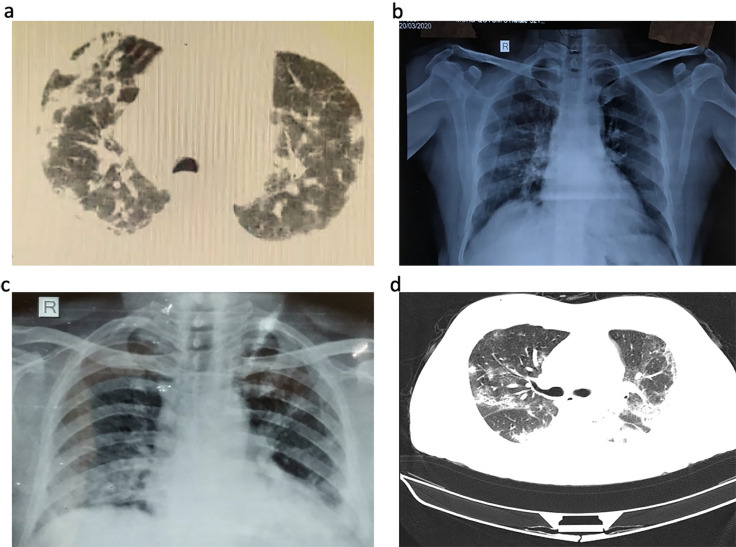
Fig 1. Chest computed tomographic, and X-ray images.
a) Chest CT images of sample ID RK100 showing chest consolidation in central areas of RT upper lobe, RT middle lobe and bilateral basal areas b) Chest X ray of sample ID KN443 showing sub pleural ground glass opacities in bilateral mid and lower zones Lt>Rt haziness c) Chest X ray of sample ID RR1191showing bilateral lower zone and left retro cardiac consolidation, Lt>Rt haziness d) multifocal bilateral ground glass opacities and patchy consolidation in a patient suffering with COVID-19.

