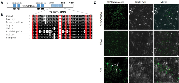
Fig. 6.
Sequence analysis of wheat E3 ubiquitin ligase [T. aestivum E3 UBQ ligase protein (TaE3UBQ)]. (A) Schematic representation of the structure of TaE3UBQ and the conserved RING finger motif using TMHMM v.2 and the CDD. (B) Sequence alignment of the C3H2C3-type RING finger conserved motif in TaE3UBQ homologues in barley (GenBank accession no. BAJ95361.1), Brachypodium (XP_003568812.1), Oryza (XP_015640584.1), maize (PWZ17186.1), Arabidopsis (NP_178156.1), millet (RLM97696.1), and sorghum (XP_002439378.1). Putative Zn2+-interacting amino acid residues are indicated in red, while conserved and non-conserved residues are highlighted in black and white, respectively. (C) GFP–TaE3UBQ fluorescent signal localizes to the cell periphery similar to the signal obtained from a plasma membrane marker pm-yk (Nelson et al., 2007) in N. benthamiana cells. The GFP fluorescent protein was found distributed throughout the cell of transformed N. benthamiana leaf cells. Scale bars=20 µm

