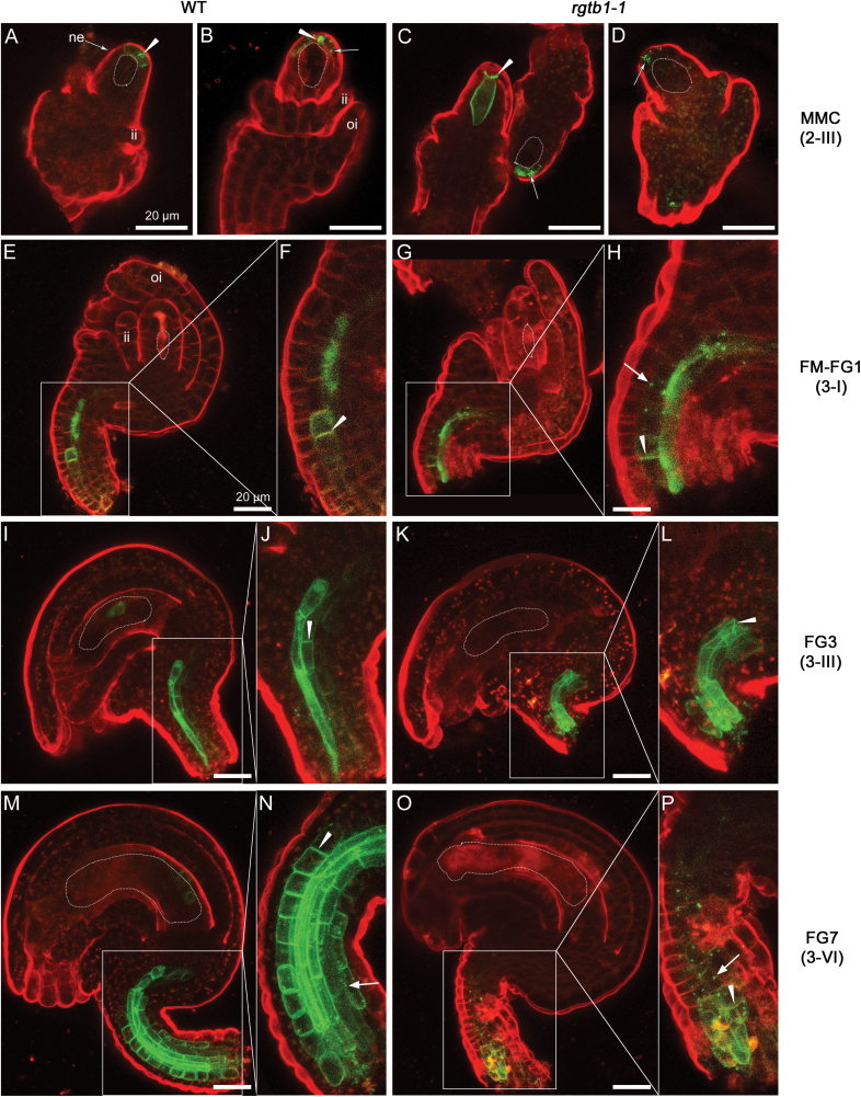
Fig. 5.
PIN3–GFP is partly internalized in the funiculus provasculature of rgtb1 ovules. pPIN3:PIN3-GFP expression in WT (A, B, E, F, I, J, M, N) and rgtb1-1 (C, D, G, H, K, L, O, P) ovules from developmental stage 2-III to 3-VI (according to Schneitz et al., 1995). PIN3–GFP localizes to MMC-adjacent membranes of the nucellus at the tip of the ovule in the WT and rgtb1-1 (A and B versus C and D); at later stages, the nucellar signal disappears in both genotypes (E–P). PIN3–GFP is also basal–polar localized in provascular cells of the funiculus in WT plants starting from the FM stage (E, F for the FM/FG1 stage; I, J for the FG3 stage; M, N for mature ovules). In rgtb1-1, PIN3–GFP is expressed in the same regions of the funiculus but is internalized to a larger extent (G, H versus E, F; K, L versus I, J; O, P versus M, N). (A–P) Confocal laser scanning microscopy, PIN1–GFP signal and FM4-64 dye fluorescence. Dotted lines highlight the MMC, FM, or FG, as appropriate for the image. Abbreviations: ne, nucellar epidermis; f, funiculus; ii, inner integument; oi, outer integument. Basal polar localization of PIN3–GFP is marked by arrowheads, and intracellular localization is marked by arrows. The left narrow image represents magnification of the area boxed in the right image. Scale bar=20 μm for all images. (This figure is available in color at JXB online.)

