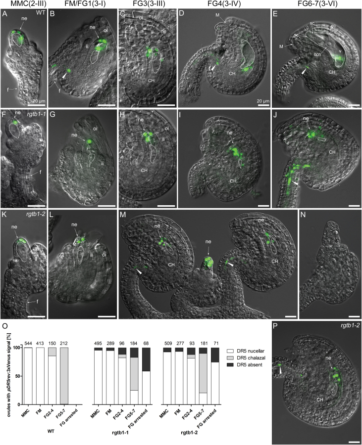
Fig. 6.
Developmentally impaired rgtb1 ovules show mislocalized response of the DR5rev:3×Venus auxin reporter. Auxin nuclear reporter DR5rev:3×Venus expression in WT (A–E), rgtb1-1 (F–J), and rgtb1-2 (K–N, P) ovules from developmental stage 2-II to 3-VI (according to Schneitz et al., 1995). DR5rev:3×Venus signal at the tip of the nucellus of WT and rgtb1 ovules at the 2-II/MMC stage (A versus F, K) until stage 3-III/FG3 (B, C versus G, H, L, M). In WT ovules from stage 3-IV/FG4 until maturity, DR5rev:3×Venus signal is localized at the chalazal part of the ovule (D, E; DR5 signal from the chalazal nucellus layer overlaps the FG layer) and the funiculus provasculature. In many rgtb1 cases, DR5rev:3×Venus signal remains localized in the micropylar pole of the ovule (for stage FG4, D versus I, M; for stage FG6, E versus J, P) and usually is also present in the funiculus. (M) rgtb1-2 ovules showing normal DR5rev:3×Venus signal localization at stage 3-III/FG3 and, in the middle, a developmentally arrested ovule expressing DR5rev:3×Venus at the tip of the nucellus. (N) Ovule arrested at an early stage of development showing no signal of DR5rev:3×Venus. (O) Fractions of ovules at different developmental stages from plants showing DR5rev:3×Venus reporter activity. Ovules were divided into three classes based on the presence of DR5rev:3×Venus signal at the top of the micropylar nucellus (white, examples in A–C, F––J, K–M, P), at the chalazal pole of the ovule in the endothelium (gray, examples in D, E), or with the signal absent (black, example in N). Ovules were counted under fluorescent and confocal microscopes. The number of ovules counted is given above each bar. (A–N, P) Epifluorescence microscopy; merged images of DIC and the DR5rev:3×Venus signal. The white dotted line highlights the MMC, FM, or FG, as appropriate for the image. Abbreviations: ii, inner integument; oi, outer integument; f, funiculus; ne, nucellar epidermis; CH, chalaza, M, micropyle; ec, egg cell; sc, synergid cell; ccn, central cell nucleus; v, central vacuole. Arrowheads point to the DR5rev:3×Venus signal in the funiculus. Scale bar=20 μm for all images. (This figure is available in color at JXB online.)

